- New stuff to read and discuss
- What patients say
- Clinic / online appointments
- Why the diagnosis of a psychosomatic illness is often a misdiagnosis
- Vascular Compression Syndromes
- Do you have questions?
- Checklist vascular compression syndromes
- Description of your symptoms
- Researchers from the Mayo Clinic confirm my concept of the Midline Congestion Syndrome
- Explanation of gender-specific differences in the clinical symptoms of abdominal vascular compression syndromes: varicocele and penile/testicular pain – their main manifestation in men.
- Varicocoele is predominantly caused by left renal vein compression
- Musculoskeletal pecularities of female puberty
- Lordosis /Swayback- Origin of many abdominal compression syndromes
- Bending of a straight vein compels its narrowing
- The lordogenetic midline congestion syndrome
- Neurological consequences of the midline congestion syndrome
- Successful treatment of a teenage girl who was unable to eat due to extreme postprandial pain and unable to walk due to spasticity in her left leg
- Severe ataxia in a young woman with severe spinal congestion – complete resolution after decompression of the left renal vein
- All compression syndromes are one: the spectrum of lordogenetic compressions
- Nutcracker-Syndrome is a misnomer! Lordogenetic left renal vein compression is a more appropriate name!
- May-Thurner-constellation (May-Thurner-syndrome, Cockett’s syndrome)
- Midline (congestion) syndrome
- Pelvic congestion syndrome
- Celiac Trunk Compression / Dunbar syndrome / MALS / Arcuate ligament syndrome
- Wilkie-Syndrome / Superior-mesenteric-artery-syndrome
- Compression of the vena cava inferior
- Evlauation of vascular compressions with the PixelFlux-method
- Connective tissue disorders predispose to multiple compressions
- Postural tachycardia syndrome (POTS) – the hemodynamic consequence of vascular compression syndromes and loose connective tissue
- Restless legs-a little known symptom of abdominal vascular compression syndromes
- Pudendal neuralgia in vascular compression syndromes
- A new sonographic sign of severe orthostatic venous pooling
- Migraine and Multiple Sclerosis
- Hemodynamic effect on cerebral perfusion in patients with multiple localised vascular compression.
- Treatment of vascular compression syndromes
- Fatal errors in the treatment of vascular compression syndromes
- Risks of stents in venous compression syndromes
- Surgical treatment of abdominal compression syndromes: The significance of hypermobility‐related disorders
- Nutcracker and May-Thurner syndrome: Decompression by extra venous tube grafting and significance of hypermobility related disorders
- Our surgical treatment of vascular compressions
- Chronic regional pain syndrome (CRPS) caused by venous compression and mechanical irritation of the coeliac plexus
- Vascular compression syndromes and other disease mechanisms I recently detected
- Kaleidoscope of instructive cases
- Venous congestion of the spinal cord may be a potential contributor to the development of paraplegia in patients with spinal muscular atrophy type III (Kugelberg-Welander disease)
- Ultrasound Diagnostics
- A breakthrough in functional sonographic diagnostic – 4D-colour Doppler sonographic flow volume measurements
- 4D-volume flow measurements of jugular and mesenteric veins
- Inauguration of the global volumetric brain perfusion measurement-a gateway for understanding of neurological symptoms
- Ultrasound focused entirely on all of your symptoms
- Ultrasound vs. X-ray
- Vascular Malformations
- Profile
- Functional colour Doppler ultrasound – how I do it
- Perfusion Measurement – PixelFlux-method
- Research
- Publications
- Nutcracker and May-Thurner syndrome: Decompression by extra venous tube grafting and significance of hypermobility related disorders
- Papers authored by Th. Scholbach
- Publications
- Inauguration of measurements of the tissue pulsatility index in renal transplants
- From nutcracker phenomenon to midline congestion syndrome and its treatment with aspirin
- First sonographic tissue perfusion measurement in renal transplants
- First sonographic bowel wall perfusion measurement in Crohn disease
- First sonographic renal tissue perfuison measurement
- First sonographic measurement of renal perfusion loss in diabetes mellitus
- PixelFlux measurements of renal tissue perfusion
- Why I prefer not to publish in journals but in the Internet
- Vessel stretching in nephroptosis – an important driver of complaints
- Publications
- Expertise
- Bornavirus Infection
- Scientific cooperation
- Cookie Policy
- Data protection
- Cookie Policy (EU)
- Impressum
Severe ataxia in a young woman with severe spinal congestion – complete resolution after decompression of the left renal vein
There are numerous consequences of a spinal cord congestion as a consequence of left renal vein and the left iliac vein compression in patients with midline congestion syndrome.
The possible numerous clinical consequences are outlined on a separate page.
I report here on a young woman developing severe vegetative symptoms, abdominal pain, convulsions and gynaecological problems due to multiple abdominal compression syndromes on the basis of a hypermobile Ehlers-Danlos syndrome.
She became wheelchair bound due to extreme ataxia of the trunk, the head and the extremities. Routine neurological diagnostics an standard imaging procedures in her home country and abroad did not reveal any treatable reason for her suffering.
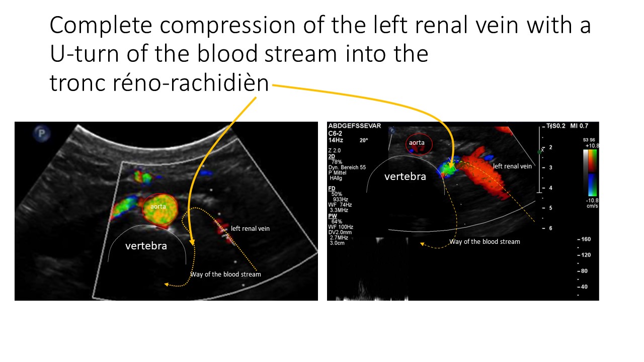
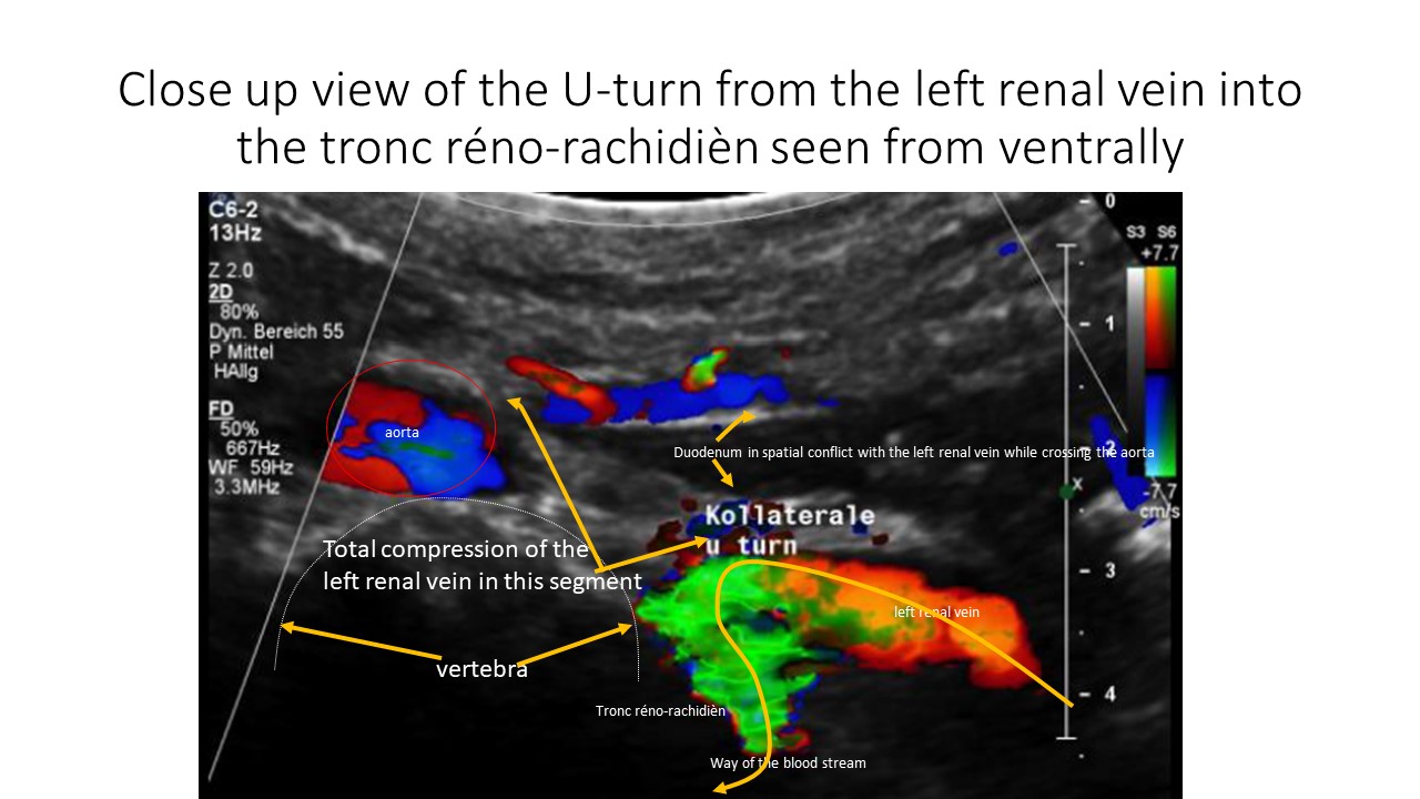
Only functional colour Doppler sonography with quantification of all relevant abdominal and pelvic vessels could demonstrate the very reason of her ordeal – a massive venous congestion of the spinal cord fed by a high flow tronc réno-rachidièn connecting the congested because completely obstructed left renal vein with the epidural plexus within the spinal canal. This produced a neurological deficit the compression and subsequent venous congestion of the spinal cord.
This video shows her miserable condition before the operation im May 2022
This video was taken in March 2023, 7,5 months after the successful decompression of the left renal vein and subsequent complete collapse of the tronc réno-rachidièn
Medical history
January 2016
- Hyperexcitability, muscle cramps, weight gain due to edema of the legs
January 2017
- Went to the emergency department because of severe muscle cramps and convulsions
- Arrhythmia, dizziness, vomiting, numbness in both hands and tremor of the trunk
- Presents several times to the emergency room of the psychiatric hospital because of general shakiness. There she was diagnosed with a mental disorder: anxiety disorder
February 2017
- Presenting to the emergency department with pain on the right side of her spine and a pollacisuria
- Taken to the psychiatric ward because of repeated generalised convulsions
- Psychiatrist refuses the diagnosis of an anxiety disorder
- Repeated convulsions
May 2017
- Was diagnosed with syndrome of intestinal bacterial overgrowth and eosinophilic oesophagitis
May 2021
- Had seen many doctors within her home country and abroad without getting a proper diagnosis and treatment for her increasing truncal instability and convulsions
- Is suffering also from abdominal pain localised under the diaphragm radiating towards both sides increasing post-prandially
- Call allannot breathe properly
- Difficulties to swallow
- Chest pain
- Always agitated
- Severe brain fog, visual disturbance, migraine, dizziness, feels like being drunk, back pain, cold and bluish hands, weak feet and hands
- Increasing fatigue
- Hyperflexibility of joints
First presentation in my functional colour Doppler sonographic clinic in June 2021
The following diagnoses had been established during the first examination:
- Pelvic congestion syndrome as a consequence of
- A May- Thurner syndrome with a venous spur
- Severe lumbar lordosis as the main reason for all vascular compression syndromes of the patient
- Complete lordogenetic compression of the left renal vein with a bypass via a
- Tronc réno-rachidièn feeding the epidural plexus and an
- Enlarged left ovarian vein transporting blood down towards the pelvis
- Substantial venous pooling in an upright position as a sign of a clinically a relevant connective-tissue disorder – in accordance with certain clinical signs of such a disorder
- Severe median arcuate ligament syndrome with typical symptoms, the main symptoms of the patient
- Ptosis of both kidneys
- Severe strangulation of blood flow towards both kidneys, mainly on the left side due to renal ptosis while being in an upright posture – this is contributing substantially to the pooling of venous blood in the pelvis and in both legs
- Heavy reduction of the circulating blood volume inside the aorta while being upright
- Double uteri


Preoperative renal parenchymal perfusion measurement in June 2021
![]()
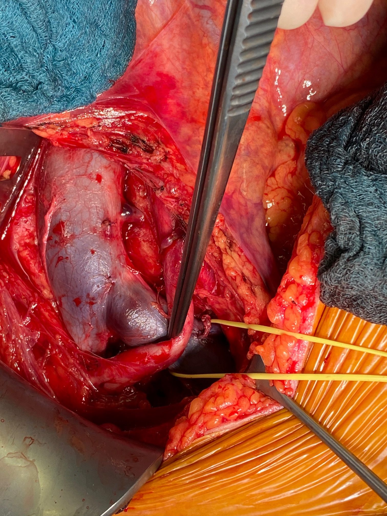
The patient was operated in June 2021 and July performing a decompression of the celiac trunc (which was complex due to heavy scar tissue found around the coeliac trunc), shielding the left common iliac vein and the left renal vein by means of a ringenforced PTFE graft.
The operation was performed by Professor Sandmann at the Clinic BelEtage in Düsseldorf (Germany). I’m very grateful for his open-minded cooperation and allownace to publish his intraoperative photos.

Liberation of the coeliac trunk after the section of massive scar tissue compressing it

Severe compression of the left common iliac vein by the crossing right common iliac artery-the sonographically diagnosed May Thurner syndrome
Degenerated wall of the left common iliac vein at the crossing site with the right common iliac artery

Liberated left renal vein with the tronc réno-rachidièn – yellow sling (blue sling: left suprarenal vein, red sling: left ovarian vein)
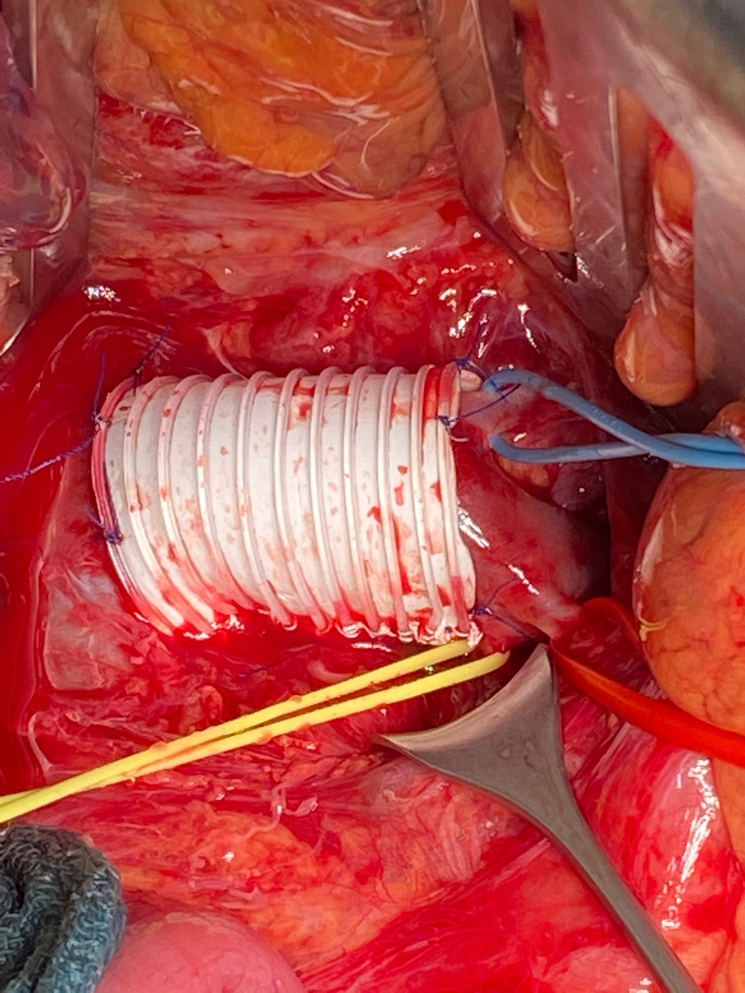
PTFE shield around the left renal vein protecting it from the compression from the aorta
Post-operative renal parenchymal perfusion measurement in November 2022 after additional decompression of the left renal vein by a PTFE-shield in July 2022
![]() In an upright posture the left to right renal perfusion ratio was 12% and is now 31%.
In an upright posture the left to right renal perfusion ratio was 12% and is now 31%.
Altogether a much better perfusion of the left kidney was achieved by the operation increasing the flow volume towards the left kidney by 87%
At the same time no blood flow across the tronc réno-rachidièn was detectable anymore whereas preoperatively about 1000 mL/min have been pushed via the tronc réno-rachidièn into the epidural plexus. This caused a substantial pressurization of the cerebral spinal fluid with a subsequent session of the spinal cord.
The reopening of the left renal vein by wrapping it with a PTFE graft made the flow across the tronc réno-rachidièn the clinical result is a complete disappearance of all neurological symptoms of the patient!
We (Prof. Sandmann and me) thank the patient for her permission to publish this report and her videos and especially for her steadfastness in not giving up hope for a cure in the face of a progressive, severe disease that couldn’t be clarified in her home country!