- El consultorio / Programación de citas en línea
- Compresiones vasculares
- ¿Tiene alguna pregunta?
- Lista de control Síndromes de compresión vascular
- Lordosis – Causa de numerosos síndromes de compresión abdominal
- Síndrome de May-Thurner/ síndrome de Cockett/ síndrome de compresión de la vena ilíaca
- Síndrome de la línea media (síndrome de congestión de la línea media)
- Síndrome de congestión pélvica (congestión de los órganos pélvicos)
- Compresión del tronco celíaco /síndrome de Dunbar/ síndrome de AMS/ síndrome del ligamento arcuato
- Síndrome de Wilkie/ síndrome de la arteria mesentérica superior
- Compresión de la vena cava inferior
- Neuralgia púdica en los síndromes de compresión vascular
- Tratamiento de los síndromes de compresión vascular
- Síndromes de compresión vascular que he detectado recientemente
- Kaleidoscope of instructive cases
- Servicios
- Ultrasonido Doppler color funcional – lo que entiendo por esto
- Medición de la circulación sanguínea – Método PixelFlux
- Investigación
- Perfil
- Infección por virus Borna
- Colaboración científica
- Cookie Policy
- Cookie Policy (EU)

Explanation of gender-specific differences in the clinical symptoms of abdominal vascular compression syndromes: varicocele and penile/testicular pain – their main manifestation in men.
Around 90% of adults who suffer from abdominal vascular compression syndromes are women. The reason for this is the greater lordosis in women than in men. This is caused by the combination of typically female anatomical features: wider hips and comparatively looser connective tissue.
However, around 10% of affected patients are male and show a different clinical picture to women.
In both sexes, the predominant abdominal vascular compressions are compression of the left renal vein and the left common iliac vein (lordogenic left renal vein compression, often incorrectly referred to as nutcracker syndrome and so-called May-Thurner syndrome).
However, the haemodynamic consequences are the same in both sexes, but the effects are different due to the different anatomy and topography (localization) of the respective genital organs.
It is helpful to know that the female and male sex organs have a corresponding embryonic origin but develop differently during embryonic and fetal life. Embryologically, the testicles correspond to the ovaries, the prostate (prostatic utricle) to the uterus, the scrotum to the labia majora and the clitoris to the penis.
However, the vascular system of these corresponding organs is basically the same in both sexes.
This means that congestion of the uterus and vagina, which is the main cause of suffering in women, corresponds to congestion of the prostate, which is often referred to as prostatitis in men.
Congestion of the external genitalia occurs somewhat less frequently in women than congestion of the internal genitalia in women. The reason for this is their relative tissue volume. The uterus is the largest single organ (in terms of its tissue mass) in the female pelvis, and therefore the uterus and vagina are the focal point of female congestion, as the congested blood seeks a path from the left to the right side of the pelvis, primarily using the relatively large vessels of the largest female pelvic organ – the uterus.
The relatively small tissue mass of the external female organs is the reason why they take second place in the development of pelvic congestion symptoms in women.
In men, on the other hand, the situation is different with regard to the size of the embryologically analogous organs and their localization outside the pelvis.
Therefore, despite the same vascular supply to the uterus and prostate, symptoms of prostatitis in abdominal congestion are rarer than symptoms of the relatively large external genital organs. The blood supply to the penis at rest and during arousal exceeds that of the clitoris.
As the congested blood seeks an auxiliary route to bypass the compression of the left iliac vein in front of the sacrum and the compression of the left renal vein in front of the upper lumbar spine, simple physical conditions lead to a different clinical picture in men and women with the same compression syndromes.
The increasing venous pressure in the two left veins follows the existing vessels. The genitals are finally drained by the uterine/prostate venous plexus, which directs the blood congested on the left side of the pelvis into the right internal iliac vein. From there, the blood flows back to the heart via the inferior vena cava to close the circulation.
However, the uterine vessels are larger than the prostate vessels, which explains the different involvement of these organs when vascular compression develops, usually during puberty. Larger vessels allow for easier uptake of additional volumes of congested blood as they offer relatively little resistance to increasing flow volumes. The primary manifestation therefore occurs in the organs with the larger vessels when all other influences are comparable.
A wider tube (blood vessel) is easier to stretch than a narrower tube. This is a well-known experience – just remember the extreme inflation pressure needed to blow up a rubber balloon (e.g., at children’s birthday parties), which drops significantly when the balloon is already slightly inflated (before rising again when the balloon is fully stretched, just before it bursts).

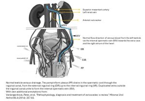
https://api-depositonce.tu-berlin.de/server/api/core/ bitstreams/1e6b0197-545f-4349-9d61-c3d333277ec6/content – with own annotations
The ovaries are always involved in the outflow obstruction of the left renal vein, as are the testicles. The most important hemodynamic difference here is the localization of these organs. The ovaries are located inside the pelvic cavity, whereas the testicles are located outside the trunk. They are subject to different environmental conditions. The pressure within the pelvis and the abdominal cavity (5 – 15 mmHg) plus the atmospheric pressure (around 760 mm Hg) resting on the body surface is significantly higher than the atmospheric pressure acting on the testicles alone. On the other hand, the hydrostatic pressure onto the testicles is somewhat higher than the pressure onto the ovaries due to the lower position of the testicles while standing upright (roughly 7 – 14 mmHg).
That is why the combination of a disturbed left kidney and left pelvic drainage is a consequence of left renal and left common iliac vein compression causes a stronger congestion of the internal female genitals and a lesser congestion of the external female genitals while in men the blood tends to prefer the circumvention via the testicles and the congestion of the prostate remains in the background.
Another key factor contributing a lot to the clinical symptoms in both sexes is gravity.
Posture has a fundamental influence on the distribution of a fluid such as blood. When lying down, gravity is directed towards the back, whereas when standing, it is directed towards the feet.
The consequences for the female and male genitals are easy to understand. Standing upright regularly leads to more severe symptoms in the genitals than lying horizontally, as the blood is drawn into the uterus by gravity (more than into the clitoris and labia), while men suffer from scrotal pain due to the posture-induced dilation of the testicular veins – the pampiniform plexus around the left testicle. This condition is known as varicocele, a painful and palpable collection of enlarged veins around the left testicle, mainly at its upper pole.
However, the penis can also become painful, and it is not uncommon for the pain to extend to the prostate if the disease persists for a longer period of time.
 The image above this from an excellent paper clearly demonstrating the broad venous connections of the may external genitals with the venous plexus of the pelvis.
Certainly, infectious prostatitis is the first assumption that urologists and general practitioners make. However, when antibiotic treatment fails, it is time to rethink and include vascular compression syndromes in the differential diagnosis.


