- New stuff to read and discuss
- What patients say
- Clinic / online appointments
- Why the diagnosis of a psychosomatic illness is often a misdiagnosis
- Vascular Compression Syndromes
- Do you have questions?
- Checklist vascular compression syndromes
- Description of your symptoms
- Researchers from the Mayo Clinic confirm my concept of the Midline Congestion Syndrome
- Explanation of gender-specific differences in the clinical symptoms of abdominal vascular compression syndromes: varicocele and penile/testicular pain – their main manifestation in men.
- Varicocoele is predominantly caused by left renal vein compression
- Musculoskeletal pecularities of female puberty
- Lordosis /Swayback- Origin of many abdominal compression syndromes
- Bending of a straight vein compels its narrowing
- The lordogenetic midline congestion syndrome
- Neurological consequences of the midline congestion syndrome
- Successful treatment of a teenage girl who was unable to eat due to extreme postprandial pain and unable to walk due to spasticity in her left leg
- Severe ataxia in a young woman with severe spinal congestion – complete resolution after decompression of the left renal vein
- All compression syndromes are one: the spectrum of lordogenetic compressions
- Nutcracker-Syndrome is a misnomer! Lordogenetic left renal vein compression is a more appropriate name!
- May-Thurner-constellation (May-Thurner-syndrome, Cockett’s syndrome)
- Midline (congestion) syndrome
- Pelvic congestion syndrome
- Celiac Trunk Compression / Dunbar syndrome / MALS / Arcuate ligament syndrome
- Wilkie-Syndrome / Superior-mesenteric-artery-syndrome
- Compression of the vena cava inferior
- Evlauation of vascular compressions with the PixelFlux-method
- Connective tissue disorders predispose to multiple compressions
- Postural tachycardia syndrome (POTS) – the hemodynamic consequence of vascular compression syndromes and loose connective tissue
- Restless legs-a little known symptom of abdominal vascular compression syndromes
- Pudendal neuralgia in vascular compression syndromes
- A new sonographic sign of severe orthostatic venous pooling
- Migraine and Multiple Sclerosis
- Hemodynamic effect on cerebral perfusion in patients with multiple localised vascular compression.
- Treatment of vascular compression syndromes
- Fatal errors in the treatment of vascular compression syndromes
- Risks of stents in venous compression syndromes
- Surgical treatment of abdominal compression syndromes: The significance of hypermobility‐related disorders
- Nutcracker and May-Thurner syndrome: Decompression by extra venous tube grafting and significance of hypermobility related disorders
- Our surgical treatment of vascular compressions
- Chronic regional pain syndrome (CRPS) caused by venous compression and mechanical irritation of the coeliac plexus
- Vascular compression syndromes I recently detected
- Kaleidoscope of instructive cases
- Ultrasound Diagnostics
- Profile
- Functional colour Doppler ultrasound – how I do it
- Perfusion Measurement – PixelFlux-method
- Research
- Publications
- Nutcracker and May-Thurner syndrome: Decompression by extra venous tube grafting and significance of hypermobility related disorders
- Papers authored by Th. Scholbach
- Publications
- Inauguration of measurements of the tissue pulsatility index in renal transplants
- From nutcracker phenomenon to midline congestion syndrome and its treatment with aspirin
- First sonographic tissue perfusion measurement in renal transplants
- First sonographic bowel wall perfusion measurement in Crohn disease
- First sonographic renal tissue perfuison measurement
- First sonographic measurement of renal perfusion loss in diabetes mellitus
- PixelFlux measurements of renal tissue perfusion
- Why I prefer not to publish in journals but in the Internet
- Vessel stretching in nephroptosis – an important driver of complaints
- Publications
- Expertise
- Bornavirus Infection
- Scientific cooperation
- Cookie Policy
- Data protection
- Cookie Policy (EU)
- Impressum

Varicocoele is predominantly caused by left renal vein compression
A varicocele is almost always the result of compression of a left renal vein
Unfortunately, it is little known that a varicocele is primarily caused by compression of the left renal vein.
This compression leads to an increase in pressure in the left renal vein and subsequently in the left spermatic vein. The increasing pressure causes dilation of the pampiniform venous plexus in the scrotum, which surrounds the testicle.
In the vast majority of cases, the left testicle is affected alone or first. The reason for this is that left renal vein compression is the most common of all abdominal compression syndromes. The pressure is transmitted directly to the left testicle. The right spermatic vein is connected to the pelvic venous plexus that surrounds the prostate, but drains directly into the inferior vena cava. Thus, after congestion of the left half of the pelvis and the left testicle, the pressure can also be transferred to the right testicle, resulting in a right varicocele.
It is therefore necessary to rule out vascular compression in patients with varicoceles. Otherwise, recurrence after ligation of the spermatic vein is very likely. The correct treatment would then be to restore the left renal outflow. Obstruction of the collateral pathway via the left spermatic vein deprives the left renal vein of its outflow pathway. The obligatory consequence is an increase in pressure and thus pain in the vascular area of the left renal vein and the formation of new collaterals, often again via the left testicle.
The following case is an instructive example:

Development of multiple enlarged and winding left spermatic veins as an attempt of the body to circumvent the compression site of the left renal vein by bypassing the blood via the left testicle towards the vena cava inferior
The video above shows a black and white ultrasound examination (B-mode ultrasound) demonstrating the large and wormlike varicose veins of the pampiniform plexus above the upper pole of the left testicle-atypical varicocele
The varicocele is however much better examined by colour Doppler ultrasound as shown in the video above. colour Doppler allows a flow quantification with the PixelFlux technique. Such measurements are the basis for an exact evaluation of the severity of the disease and moreover can precisely describe the effect of any treatment.

Exact quantification of the blood flow in the pampiniform plexus and the testicle left and right by the PixelFlux measurement
With the PixelFlux measurement, it can be precisely calculated that the flow volume in the left pampiniform plexus is 7 times higher than in the right, while the blood flow in the left testicle is 3 times higher than in the right.
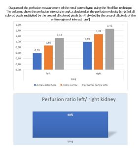
Not only allows PixelFlux to quantify the varicocele itself but also the underlying left renal vein compression which is demonstrated in the following diagram:
The renal perfusion measurement with the PixelFlux technique shows that the high pressure in the left renal vein cannot be compensated by the circumvention of blood across the left testicle since even though the left kidney receives only 68% of the blood flow volume of the right kidney. Thus, not only the left testicle but also the left kidney is suffering.
Normal right testicle of the same patient in the video above.
Accordingly, the blood flow within the pampiniform plexus of the right testicle is significantly lower.


