- New stuff to read and discuss
- What patients say
- Clinic / online appointments
- Why the diagnosis of a psychosomatic illness is often a misdiagnosis
- Vascular Compression Syndromes
- Do you have questions?
- Checklist vascular compression syndromes
- Description of your symptoms
- Researchers from the Mayo Clinic confirm my concept of the Midline Congestion Syndrome
- Explanation of gender-specific differences in the clinical symptoms of abdominal vascular compression syndromes: varicocele and penile/testicular pain – their main manifestation in men.
- Varicocoele is predominantly caused by left renal vein compression
- Musculoskeletal pecularities of female puberty
- Lordosis /Swayback- Origin of many abdominal compression syndromes
- Bending of a straight vein compels its narrowing
- The lordogenetic midline congestion syndrome
- Neurological consequences of the midline congestion syndrome
- Successful treatment of a teenage girl who was unable to eat due to extreme postprandial pain and unable to walk due to spasticity in her left leg
- Severe ataxia in a young woman with severe spinal congestion – complete resolution after decompression of the left renal vein
- All compression syndromes are one: the spectrum of lordogenetic compressions
- Nutcracker-Syndrome is a misnomer! Lordogenetic left renal vein compression is a more appropriate name!
- May-Thurner-constellation (May-Thurner-syndrome, Cockett’s syndrome)
- Midline (congestion) syndrome
- Pelvic congestion syndrome
- Celiac Trunk Compression / Dunbar syndrome / MALS / Arcuate ligament syndrome
- Wilkie-Syndrome / Superior-mesenteric-artery-syndrome
- Compression of the vena cava inferior
- Evlauation of vascular compressions with the PixelFlux-method
- Connective tissue disorders predispose to multiple compressions
- Postural tachycardia syndrome (POTS) – the hemodynamic consequence of vascular compression syndromes and loose connective tissue
- Restless legs-a little known symptom of abdominal vascular compression syndromes
- Pudendal neuralgia in vascular compression syndromes
- A new sonographic sign of severe orthostatic venous pooling
- Migraine and Multiple Sclerosis
- Hemodynamic effect on cerebral perfusion in patients with multiple localised vascular compression.
- Treatment of vascular compression syndromes
- Fatal errors in the treatment of vascular compression syndromes
- Risks of stents in venous compression syndromes
- Surgical treatment of abdominal compression syndromes: The significance of hypermobility‐related disorders
- Nutcracker and May-Thurner syndrome: Decompression by extra venous tube grafting and significance of hypermobility related disorders
- Our surgical treatment of vascular compressions
- Chronic regional pain syndrome (CRPS) caused by venous compression and mechanical irritation of the coeliac plexus
- Vascular compression syndromes and other disease mechanisms I recently detected
- Kaleidoscope of instructive cases
- Venous congestion of the spinal cord may be a potential contributor to the development of paraplegia in patients with spinal muscular atrophy type III (Kugelberg-Welander disease)
- Ultrasound Diagnostics
- A breakthrough in functional sonographic diagnostic – 4D-colour Doppler sonographic flow volume measurements
- 4D-volume flow measurements of jugular and mesenteric veins
- Inauguration of the global volumetric brain perfusion measurement-a gateway for understanding of neurological symptoms
- Ultrasound focused entirely on all of your symptoms
- Ultrasound vs. X-ray
- Vascular Malformations
- Profile
- Functional colour Doppler ultrasound – how I do it
- Perfusion Measurement – PixelFlux-method
- Research
- Publications
- Nutcracker and May-Thurner syndrome: Decompression by extra venous tube grafting and significance of hypermobility related disorders
- Papers authored by Th. Scholbach
- Publications
- Inauguration of measurements of the tissue pulsatility index in renal transplants
- From nutcracker phenomenon to midline congestion syndrome and its treatment with aspirin
- First sonographic tissue perfusion measurement in renal transplants
- First sonographic bowel wall perfusion measurement in Crohn disease
- First sonographic renal tissue perfuison measurement
- First sonographic measurement of renal perfusion loss in diabetes mellitus
- PixelFlux measurements of renal tissue perfusion
- Why I prefer not to publish in journals but in the Internet
- Vessel stretching in nephroptosis – an important driver of complaints
- Publications
- Expertise
- Bornavirus Infection
- Scientific cooperation
- Cookie Policy
- Data protection
- Cookie Policy (EU)
- Impressum
Genital and sexual symptoms of vascular compression syndromes – patient with ulcer of labia minora
In venous vascular compression syndromes, congestion of venous blood in the lower half of the body develops.
The constriction of the left iliac vein at the May Thurner point, the crossing with the right iliac artery, which is usually found slightly below the navel and to the right of it, hinders an undisturbed return flow of blood from the legs and pelvic organs and from the gluteal muscles back to the heart. The veins in the pelvis and legs and along the sciatic nerve dilate and a feeling of tightness in the pelvis and legs and later increasingly severe pain here and along the sciatic nerve result.
It is also not uncommon for unpleasant, unwanted, and unprovoked sexual arousal to develop, as well as for missensations such as burning, formication, buzzing, and similar symptoms to develop.
The genital organs are particularly affected by such blood stasis because they are located at the lowest point of the trunk. When the body is in an upright position, sitting or standing, the blood, following the force of gravity, therefore accumulates especially in the area of the internal and external genital organs.
These symptoms are particularly aggravated when other vascular compression syndromes are also present, which is a regular occurrence.
Compression of the left renal vein leads to an increase in venous congestion in the pelvis, since the left ovarian vein and the left testicular vein, respectively, drain blood from the congested renal vein into the pelvis.
Not infrequently, there is also compression of the right iliac vein on the L4/L5 disc, as well as compression of the inferior vena cava.
Compression of the inferior vena cava occurs with pronounced lordosis of the lumbar spine mostly in the upper abdominal cavity, only a few centimeters below the right costal arch. Here, with the mostly flat thorax of the patients, the liver presses anteriorly and superiorly on the inferior vena cava, which lies on a firm abutment on the cranial slope of the lordotic spinal curve and therefore cannot avoid the pressure of the liver, especially in standing position when the liver descends.
The combination of these venous compressions is the rule and not the exception. It is therefore necessary to measure all possible vascular compression syndromes in the abdomen, on the one hand to draw the correct diagnostic conclusions, and on the other hand to be able to make a successful correction.
Often, the symptoms in the genitals are aggravated when sitting or standing.
In addition to the general effect of gravity, which draws blood to the lowest point of the trunk or legs when the trunk is in an upright position, the change in renal blood flow when the trunk is in an upright position plays a major role in the development of these symptoms.
Since the kidneys receive a particularly abundant supply of blood – one kidney, for example, receives much more blood than an entire leg – the kidneys’ blood intake significantly affects blood flow to the pelvis. As soon as and if kidney blood flow deteriorates while the trunk is upright, blood, coming from the heart, flows past the openings of the renal arteries and pours into the pelvis. If the above-mentioned venous vascular compression syndromes are present, this very quickly results in enormous blood filling in the pelvis and legs, which particularly affects the genitals, since they are located at the lowest point of the trunk when the trunk is in an upright posture and the blood collects there first.
A drastic decrease in renal blood flow is not uncommon in patients with connective tissue weakness, in whom the kidneys slide downward while sitting or standing and throttle their own blood supply by pulling on the renal vessels.
Unfortunately, this cannot be visualized with ordinary imaging techniques. Computer tomography and MRI are unable to detect these functional changes because, on the one hand, they are only performed in a lying body position and, on the other hand, they are not able to determine the blood supply to the kidney in routine operation.
With the PixelFlux technique, it is possible to detect the reduction in blood flow to the kidneys within a few seconds. Its extent can also be measured accurately. In addition, during the functional examination in the supine and standing position, the blood flow in the entire abdominal cavity can be determined by quantifying the flow volume in the aorta. Even the stroke volume of the heart can be determined in a straightforward manner by sonographically measuring the filling of the left ventricle during the abdominal examination.
In this way, it is possible to determine exactly how much additional blood accumulates in the pelvis when the body changes position from horizontal to vertical. In this way, the position-dependent pain in the genitals but also in the legs can be correlated precisely with objective measurements.
In the following patient, there had already been severe pain in the external genitals for a long time and sexual intercourse was not possible because of the severe vaginal pain.
After a vaginal gynecological examination with specula (examination spatulas), a large painful ulcer developed on the left labia minora. Unaware of the massive venous congestion of the patient’s genitalia, the attending physicians were unable to find the cause of this painful ulcer, as all standard examinations to detect sexually transmitted diseases remained negative.
Significant swelling of the left labium developed. After the ulcer slowly subsided, additional ulcers developed on the posterior commissure, the site of the posterior union of the labia.
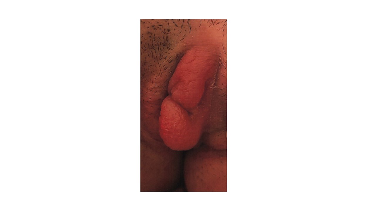
Massive edema of the small left labium
PixelFlux examination of the external genitalia demonstrated that significant left-sided venous congestion of the labia had resulted in severe edema of the tissue.

Edema of the left labia on color Doppler ultrasound
Pixelflux measurement of the congestion of the labia.
The maximum tissue perfusion of the labia measured under standardized conditions (cm/s * cm² of the perfused area of the ROI /cm² of the ROI) is:
| Small labia | |
| left | 0,243 |
| right | 0,167 |
The significant lateral difference in blood flow to the labia could be visualized and determined in detail. It thus became clear that the particular left-sided venous congestion typical of May-Thurner syndrome was the cause of the particular sensitivity of the swollen and poorly perfused tissue. Venous congestion regularly leads to an insufficient supply of oxygen to the tissues, in addition to the repair of the tissues, which leads to a long-lasting inflammation. These changes make the tissue particularly sensitive to pressure from the examination spatula during vaginal gynecological examination. By even small pressure then the tissue can be easily destroyed, which had led to tissue destruction, necrosis and thus a large ulcer.

Ulcer of the left labia minora over time