- 超音波診断外来
- 血管圧迫症候群
- 血管圧迫症候群の治療
- 最近発見された血管圧迫症候群
- 万華鏡のように参考になる事例が多い
- 機能性カラードップラー超音波検査 – どのようにして行うか
- 血流測定 – PixelFlux法
- 研究
- 専門知識
- ボルナウイルス感染症
- クッキーポリシー
- 医学的な説明に関する注意事項
- 個人情報保護方針
- Cookie Policy (EU)

Unbearable pain in the right kidney when standing or sitting, as well as persistent pelvic pain despite stenting of the left iliac vein. This can be explained by two detrimental stent mechanisms that were unknown until now.
This patient had an intravenous stent inserted into the left common iliac vein to treat May–Thurner syndrome. Following the procedure, she developed unbearable pain in her right kidney while being upright preventing her from standing or sitting. While the standard examination while lying supine did not reveal any cause of this specific symptom, a repeat examination while standing demonstrated severe right-sided orthostatic renal nephroptosis of 13.2 cm.
While standing, the intravenous stent tilted ventrally and cranially, touching and lacerating the right kidney as it sank downwards. The sharp, triangular tips of the stent were poking 2 mm into the renal parenchyma, causing unbearable pain. This explains why the pain persisted, albeit less severely, when lying horizontally, since repeated wounding of the kidney when performing daily activities in an upright posture may have caused a chronic wound that was painful at all times, but which caused unbearable pain when the stent actually penetrated the kidney surface while standing or sitting.
The following medical video illustrates the situation.
This is another example of the detrimental effect of intravenous stents in patients with vascular compression syndromes of the abdomen and pelvis.
Additionally, the patient experienced persistent pelvic pain, which was due to persistent venous congestion of the pelvic organs demonstrated by the PixelFlux technique.
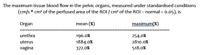
Exact description of the degree of pelvic congestion by multiple PixelFlux measurements of the tissue perfusion of relevant pelvic organs
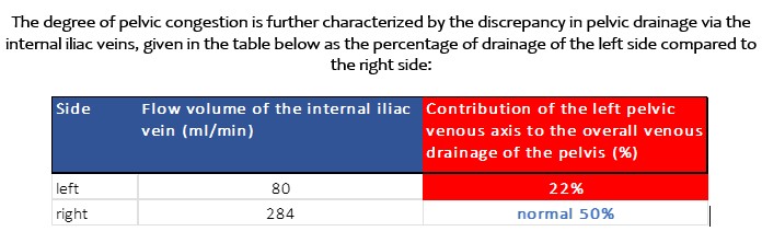
Direct flow measurements of both internal iliac veins demonstrated the degree of the insufficient drainage of the left pelvic hemisphere
This congestion was a consequence of the stent’s insufficiency. Despite the stent’s uncompromised 12 mm diameter, blood flow could not enter the stent completely. A comparison of the flow volumes of the left internal and external iliac veins revealed a flow volume of 335 ml/min. However, within the stent, the flow volume was only 214 ml/min. Therefore, the stent could only transport 64% of the blood volume that attempted to enter it.
This was due to the angulation of the stent relative to the native vein. Due to its angulation, the stent obstructed the vein and, in addition, the rigid wall of the stent transformed the kinetic energy of the bloodstream into warmth, thus reducing the energy available to propel the bloodstream.
This is in stark contrast to a native vein, which is elastic. Its elastic wall allows the vein to distend during pressure changes, which occur constantly during breathing, movement, bending the trunk and using the leg muscles, thus increasing venous return. Such pressure changes cause the elastic vein to distend momentarily, and as soon as the pressure drops, the elastic wall contracts, thus propagating the blood stream. Thus, kinetic energy is transferred into elastic force, which stores the kinetic energy of the bloodstream and releases it when the vein contracts again. This mechanism is unavailable to a rigid metal stent, which is why there is constant energy loss when the stent is angulated and the blood stream is repeatedly reflected off the stent wall.
The stent can be compared to a corridor. If you try to throw a ball through a corridor, you will succeed as long as the ball does not touch the corridor walls. This would be similar to a stent that is completely aligned with the entering vein. However, if there is an angle between the entering vein and the stent, the situation changes fundamentally. It would be similar to trying to throw a ball obliquely against the corridor wall. The ball would be reflected towards the opposite wall, losing its kinetic energy in the process. The ball will stop moving after a few bounces, meaning it will not make it to the end of the corridor.
The same situation applies to a curved stent or a stent that is not aligned with the entering or exiting vein. This is the rule in vascular compression syndromes, since compressed veins always run in a curve due to being lifted by the spine. Therefore, the angle of the stent with the vein always reduces the transfer of blood across the stent compared to a normal vein. This is why even a well-sized and open stent may become insufficient. This mechanism is not widely known and is rarely considered when patients complain of persistent symptoms despite stenting of the vein.