- 超音波診断外来
- 血管圧迫症候群
- 血管圧迫症候群の治療
- 最近発見された血管圧迫症候群
- 万華鏡のように参考になる事例が多い
- 機能性カラードップラー超音波検査 – どのようにして行うか
- 血流測定 – PixelFlux法
- 研究
- 専門知識
- ボルナウイルス感染症
- クッキーポリシー
- 医学的な説明に関する注意事項
- 個人情報保護方針
- Cookie Policy (EU)
Successful treatment of severe post-prandial upper abdominal pain due to pancreatic congestion in a patient with high-grade compression of the splenic vein
A 24-years old female patient had been operated to decompress the initially severely compressed left renal vein. Postoperatively she developed a compression of the splenic vein by the coeliac trunk against the prepyloric antrum of the stomach and a progressive left common iliac vein compression.
Her main complaints were unbearable post-prandial epigastric pain (severity 9/10) , back pain and she was unable to stand for longer periods.
For the past month, the patient has had 6 episodes of increasing pain located below the left ribcage and radiating to the spine on the left side. In addition, she experienced more pain while working and a different type of pain while eating. As a result, she lost about 10 pounds in the last 3 months by avoiding the uptake of food to reduce the pain.
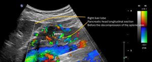
Prior to the operation the patient was suffering from severe post-prandially accentuated epigastric pain which could be referred to the massive congestion of the pancreas. The following videos demonstrate the hypervascular pancreatic head and pancreatic tail.
Preoperative sweep across the pancreas showing a massive vascularisation of the entire organ due to its involvement into the collateral pathway of the severely compressed splenic vein
Video showing the congested pancreatic head with a network of multiple intertwined congested veins. The extent of the congestion can only be measured with the PixelFlux technique-see below
Detailed post-operative view of the pancreatic head. The decompression of the splenic vein renders the collateral network in the pancreas unnecessary which thus disappears due to its collaps.
Vide showing the pancreatic tail after the decompression of the splenic vein-now no blood vessels can be depicted anymore inside the pancreas- a proof of the former involvement of the pancreas in the collateral pathway of the compressed splenic vein
Close up view of the former compression site of the splenic vein at the crossing with the coeliac trunk after its decompression
Worm- like intertwining enlarged parenchymal veins inside the head of the pancreas due to its involvement in the collateral pathway of the compressed splenic vein prior to the surgical decompression

After surgical decompression of the splenic vein the venous collateral network inside the pancreatic head completely disappeared. A clear. the pancreas was involved in the collateral pathway of the compressed splenic vein.
The only way to quantify the extent of the collateralisation of a parenchymal organ by means of ultrasound-which is obviously the most patient-friendly imaging technique-is the PixelFlux measurement:
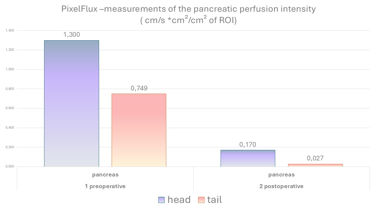
The PixelFlux measurement can quantify the extent of this congestion and also allow a comparison to the profound improvement with a complete resolution of this congestion after the decompression of the splenic vein. Obviously, the congestion of the pancreas was an effect of the involvement of the pancreas into the collateral pathway of the congested splenic vein.
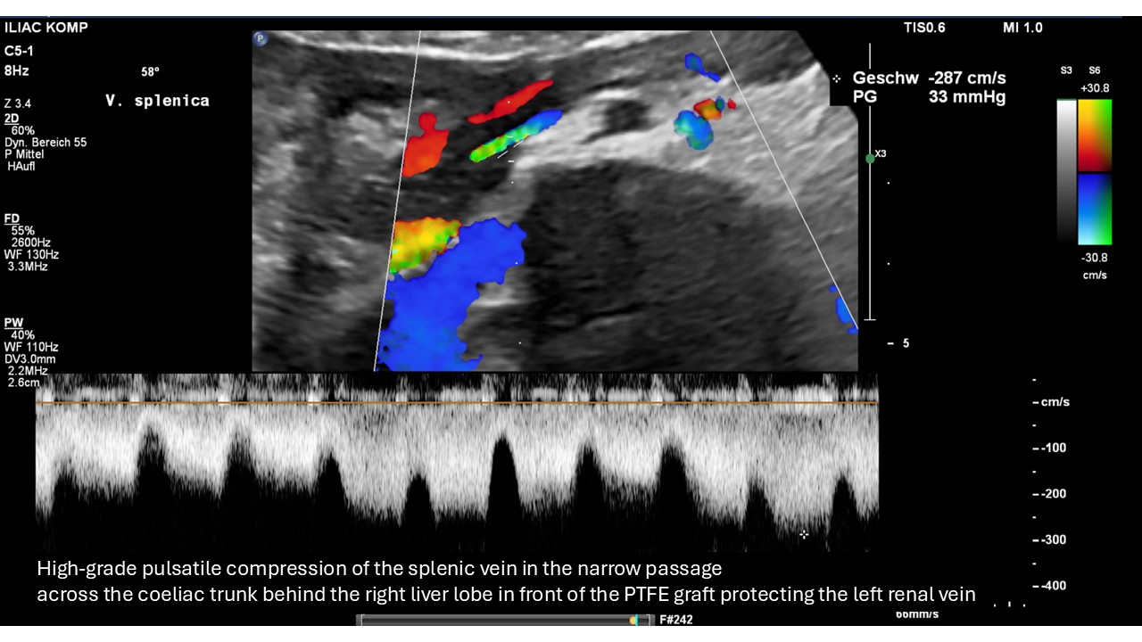
Strong flow acceleration within the splenic vein as a sign of its severe compression by the coeliac trunk (small round structure in the upper Centre of the image). The pulsatile nature of the flow acceleration is the proof that an artery-here the coeliac trunk-is the compressing structure.

Close-up view of the compression of the splenic vein the coeliac trunk.

Ultrasound B-mode image demonstrating the compression of the splenic vein with the surrounding structures which narrow the space for the splenic vein thus preparing its compression.

Spatial relationship of the PTFE graft protecting the formerly compressed left renal vein and the splenic vein showing that graft itself is not compressing the splenic vein.

After the surgical decompression of the splenic vein the flow velocity drops to the half of the preoperative values demonstrating the significant reduction of the

Detailed view of the now apparently vessel-free pancreatic tail after the are decompression of the splenic vein rendering the collateral pathway via the pancreas unnecessary.