- संवहनी संपीड़न सिंड्रोम
- क्या आपका कोई सवाल है?
- चेकलिस्ट संवहनी संपीड़न सिंड्रोम
- महिला यौवन की मस्कुलोस्केलेटल विशेषताएं
- लॉर्डोसिस – कई उदर संपीड़न सिंड्रोम का कारण
- नटक्रैकर सिंड्रोम एक मिथ्या नाम है
- मे-थर्नर नक्षत्र / मे-थर्नर सिंड्रोम / कॉकटेल सिंड्रोम / वेना इलियाक संपीड़न सिंड्रोम
- पेल्विन्स कंजेशन सिंड्रोम (श्रोणि अंगों का जमाव)
- ट्रंकस कोएलियाकस संपीड़न / डनबर सिंड्रोम / MALS / लिगामेंट आर्किकेट सिंड्रोम
- विल्की सिंड्रोम / बेहतर मेसेन्टेरिक धमनी सिंड्रोम
- PixelFlux तकनीक का उपयोग करके संवहनी संपीड़न सिंड्रोम की मात्रा
- संवहनी संपीड़न सिंड्रोम में पुदाल तंत्रिकाशूल
- संवहनी संपीड़न सिंड्रोम का उपचार
- संवहनी संपीड़न सिंड्रोम जो मैंने हाल ही में खोजा है
- Kaleidoscope of instructive cases
- कार्यात्मक रंग डॉपलर अल्ट्रासाउंड – जैसा कि मैं इसे समझता हूं
- अनुसंधान
- विशेषज्ञता
- वैज्ञानिक सहयोग
- Cookie Policy
- Cookie Policy (EU)

Abdominal pain due to liver compression in slipping rib syndrome in an EDS patient
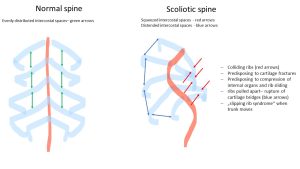
Patients with Ehlers-Danlos syndrome and other connective tissue disorders suffer from the reduced strength of connective tissue structures. As a result, they often develop joint dislocations or subluxations, overstretched ligaments, excessive curvatures of the spine such as an unusually pronounced lordosis of the lumbar spine (forward curvature), kyphosis (backward curvature-hump formation) of the thorax, or scoliosis (a sideways curvature of the spine). Often, increased lordosis and kyphosis precede scoliosis. The bending of the spine is favored by the overstretchable ligaments of the spine.
However, cartilaginous structures that naturally contain a great deal of connective tissue also often exhibit decreased stability. These include not only the cartilaginous articular surfaces but also, for example, the cartilage bridges that connect the anterior ends of the ribs to each other and to the sternum.
In particular, broad cartilage ridges form between the 8th and 10th ribs, which can break more easily in patients with Ehlers-Danlos syndrome, so that the affected ribs are then much more mobile than usual and the stability of the rib cage is reduced as a result.
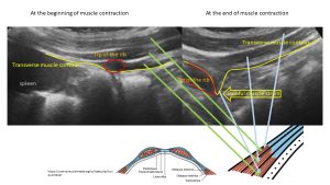
It then happens that during certain movements that involve tightening of the abdominal muscles (straightening from a supine position, twisting movements of the trunk), these freed ribs are pressed inward and slide under adjacent ribs. This can sometimes be felt by the affected persons themselves.
Directly at the lower edge of each rib, in addition to an intercostal artery and an intercostal vein, there is also a very pain-sensitive intercostal nerve, which is responsible for activating the intercostal muscles.
If such a broken out rib slides under other ribs, this can lead to very painful irritation of the intercostal nerve of the rib above and to a painful strain of the muscles of the abdominal and thoracic wall. In addition, the broken rib may press on internal organs such as the liver, causing pain and even organ damage (elevation of liver enzymes in the blood).
This clinical picture, which is rarely considered, can be reliably detected by means of a functional ultrasound examination and is called slipping-rib syndrome.
The following pictures illustrate the mentioned changes in the case of a young woman with Ehlers-Danlos syndrome, who suffered from concomitant vascular compression syndromes but additionally from thoracic and upper abdominal pain, which was dependent on certain movements and postures. She also had unexplained elevations of liver enzymes in her blood.
More can be found here: Cartilaginous compression of the liver – clinical and ultrasonographic aspects
(link provided by an enthusiastic patient)
The following video shows the movement of the 8th/9th and 10th ribs on the right side converging as the patient tenses her abdominal muscles. As this occurs, the tips of the ribs converge and the 9th rib slides under the 8th rib, which is associated with severe pain in the right lower thorax and right upper abdomen, respectively.
These images show the significant compression of the liver by the ribs pushing inward, which have lost their cohesion as the cartilage bridge is broken.
Loosening of ribs has also occurred on the left side. Since the liver is not located here, but the smaller spleen, the mobile rib pushes inward into the abdominal cavity and painfully stretches the muscles of the abdominal wall.
Treatment of the condition consists of surgical stabilization of the ribs or shortening them.


